Blood Vessels Labeled - Blood Vessel Man Model Youtube / Name the blood vessel labeled 'b'.
bymamafairrow•
0
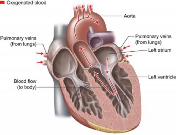
Blood Vessels Labeled - Blood Vessel Man Model Youtube / Name the blood vessel labeled 'b'.. Arteries transport blood away from the heart. Labeled arm showing the antecubital veins / normal blood pressure is 120/80. That being said, all arterial blood delivered to this region comes via branches of the abdominal aorta, and all venous blood eventually finds its way back to. Label the major blood vessels of the pulmonary and systemic circulations; Veins (in blue) are the blood vessels that return blood to the heart.
Blood vessels 11p image quiz. The vessels that carry blood away from the heart are called arteries, and their very small branches are arterioles. Blood cells by descartes 48,739 plays 9p image quiz. Labeled arm showing the antecubital veins / normal blood pressure is 120/80. The common cartoid artery extends from the brachiocephalic artery.
How The Heart Blood Vessels Work Heart Vascular Institute Temple Health from www.templehealth.org What is the name of blood vessel b? Eventually, the smallest arteries, vessels called arterioles, further branch into tiny capillaries, where nutrients and wastes are exchanged. The three major types of blood vessels: Blood cells by descartes 48,739 plays 9p image quiz. Arteries transport blood away from the heart. Veins return blood back toward the heart. Blood vessels 11p image quiz. Blood vessels prepared by dr.
It is the innermost and thinnest layer of arteries and veins, which have a direct contact with the blood flow.;
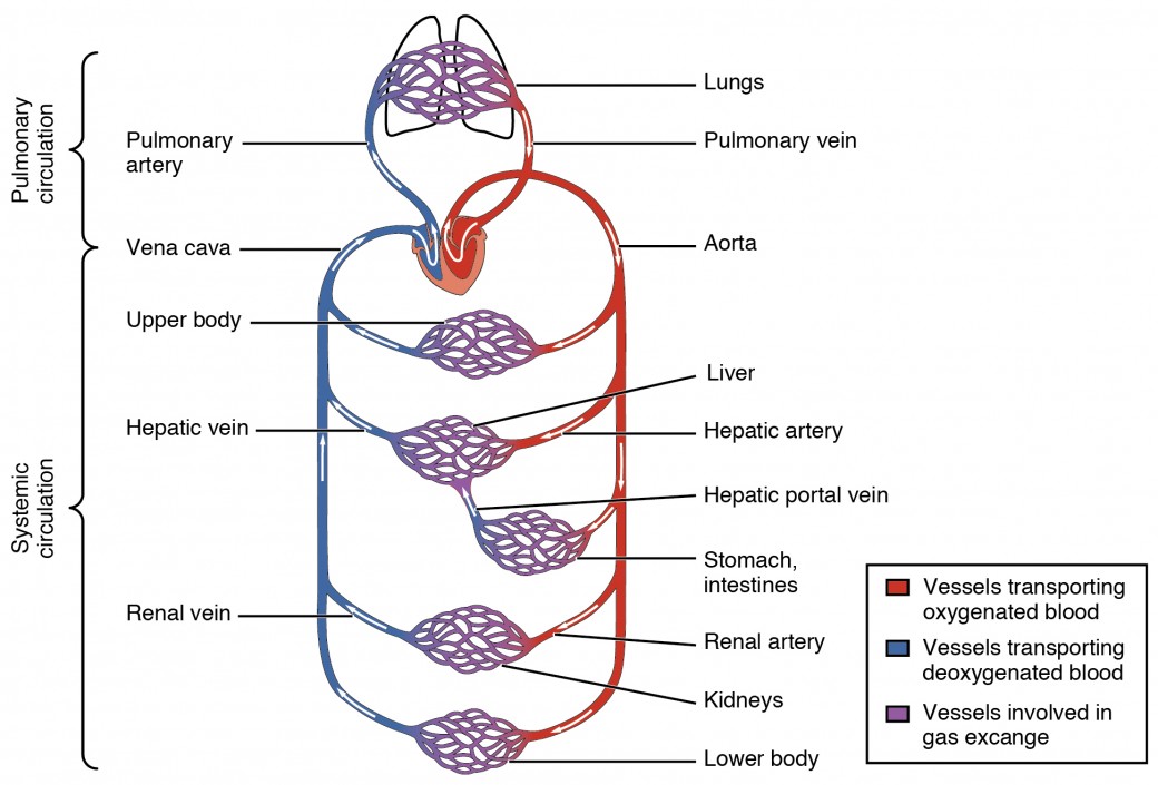
Arteries (in red) are the blood vessels that deliver blood to the body. Human heart labeling 27p image quiz. The vessels that carry blood away from the heart are called arteries, and their very small branches are arterioles. The three major types of blood vessels: The venules and veins returning blood to the heart. Deep veins, located in the center of the leg near the leg bones, are enclosed by muscle. Deoxygenated blood from the peripheral veins is transported back to the heart from capillaries, to venules, to veins, to the right side of the heart, and then. Blood vessels of the abdomen and pelvis. Navigate to the cardiovascular system area in the following pal 3.0 module: •formed where capillaries unite • extremely porous 1) venules: The common cartoid artery extends from the brachiocephalic artery. It extends on each side of the neck and divides at the level of the larynx into two branches: Both arteries and veins consist of three layers.
Liver anatomy blood supply 19 photos of the liver anatomy blood supply anatomical location of liver, blood vessels that carry blood to the liver, dual blood supply to liver, functional anatomy of liver, liver and its functions, liver on the human body, normal anatomy of the liver, position of liver, inner body, anatomical location of … As the abdomen and pelvis contain the majority of internal organs, these regions need to be supplied by an extensive network of arteries and veins. Arm blood vessels labeled : Describe the development of blood vessels and fetal circulation; Blood vessel structure and function.
Structure And Function Of Blood Vessels Anatomy And Physiology Ii from s3-us-west-2.amazonaws.com Eventually, the smallest arteries, vessels called arterioles, further branch into tiny capillaries, where nutrients and wastes are exchanged. Name the blood vessels labeled 'e'. The adventitia or outer layer which provides structural support and shape to the vessel Name the blood vessel labeled 'd'. That being said, all arterial blood delivered to this region comes via branches of the abdominal aorta, and all venous blood eventually finds its way back to. Eventually, the smallest arteries, vessels called arterioles, further branch into tiny capillaries, where nutrients and wastes are exchanged, and then combine with other vessels that exit capillaries to form venules, small blood vessels that carry blood to a vein, a larger blood vessel that returns blood to the heart. Liver anatomy blood supply 19 photos of the liver anatomy blood supply anatomical location of liver, blood vessels that carry blood to the liver, dual blood supply to liver, functional anatomy of liver, liver and its functions, liver on the human body, normal anatomy of the liver, position of liver, inner body, anatomical location of … Vessels transport nutrients to organs/tissues and to transport wastes away from organs/tissues in the blood.
The vessels that carry blood away from the heart are called arteries, and their very small branches are arterioles.
Blood vessel labeling 9p image quiz. Structure & function of blood vessels. Blood vessels of the abdomen and pelvis. Blood vessels 11p image quiz. Name the blood vessel labeled 'c'. Identify and describe the hepatic portal system; This article lists a series of labeled imaging anatomy cases by system and modality. Deoxygenated blood from the peripheral veins is transported back to the heart from capillaries, to venules, to veins, to the right side of the heart, and then. The superior vena cava is not labeled in figure 7.4. The common cartoid artery extends from the brachiocephalic artery. Vessels transport nutrients to organs/tissues and to transport wastes away from organs/tissues in the blood. Name the blood vessel labeled 'b'. A primary purpose and significant role of the vasculature is its participation in oxygenating the body.
The word vascular, meaning relating to the blood vessels, is derived from the latin vas, meaning vessel. It is present adjacent to the tunica media and is composed of collagen and. Name the blood vessel labeled 'd'. It is the innermost and thinnest layer of arteries and veins, which have a direct contact with the blood flow.; The superior vena cava is not labeled in figure 7.4.
Art Labeling Quiz from wps.pearsoned.com The word vascular, meaning relating to the blood vessels, is derived from the latin vas, meaning vessel. It is present adjacent to the tunica media and is composed of collagen and. Capillaries come together to form venules, small blood vessels that carry blood to a vein, a larger blood vessel that returns blood to the heart. The inferior vena cava is labeled in the figure below. Blood vessel labeling 15p image quiz. Blood vessels of the head and neck. It is the innermost and thinnest layer of arteries and veins, which have a direct contact with the blood flow.; Eventually, the smallest arteries, vessels called arterioles, further branch into tiny capillaries, where nutrients and wastes are exchanged, and then combine with other vessels that exit capillaries to form venules, small blood vessels that carry blood to a vein, a larger blood vessel that returns blood to the heart.
Vessel networks deliver blood to all tissues in a directed and regulated manner.
Blood vessel labeling 9p image quiz. The thick outermost layer of a vessel (tunica adventitia or tunica externa ) is made of connective tissue. A primary purpose and significant role of the vasculature is its participation in oxygenating the body. Vessels transport nutrients to organs/tissues and to transport wastes away from organs/tissues in the blood. •formed where capillaries unite • extremely porous 1) venules: Name the blood vessels labeled 'e'. External veins and arteries of the heart ec by mrsdohm 64,784 plays 8p image quiz. Blood vessels consist of arteries, arterioles, capillaries, venules, and veins. Blood vessels 11p image quiz. It is the innermost and thinnest layer of arteries and veins, which have a direct contact with the blood flow.; Arteries can be classified based on the abundance of elastic fibres present in the walls. Both arteries and veins consist of three layers. The adventitia or outer layer which provides structural support and shape to the vessel